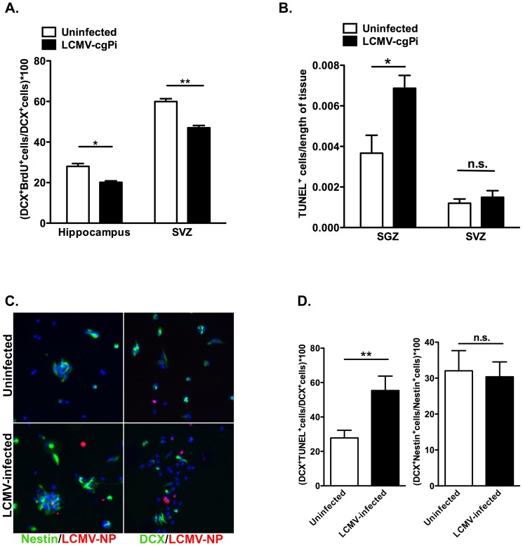
Figure 3. Proliferation and survival of neural progenitor cells are affected in LCMV-cgPi mice.
In age-matched 6-week old mice, (A) analysis of neural progenitor cells using flow cytometry revealed lower percentages of BrdU-labeled neuroblasts in the hippocampus and SVZ of LCMV-cgPi mice. Data points are shown with mean ± SEM, N≥4 mice; *p<0.05, **p<0.01. (B) Quantitative IHC analysis revealed greater numbers of TUNEL+ cells in the SGZ of LCMV-cgPi mice. Data points are shown with mean ± SEM, N = 3 mice. (C) Neurospheres were infected with LCMV, and growth factors were removed to induce differentiation. Representative ICC images show Nestin+ transit amplifying cells and DCX+ neuroblasts in neurosphere cultures at 12 hours following withdrawal of growth factors. (D) Quantitative ICC analysis revealed increased numbers of TUNEL+Nestin+ cells in the LCMV-infected cell cultures. Data points are shown with mean ± SEM, N = 3 wells.

