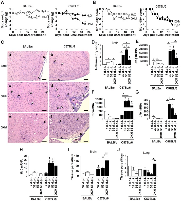
Figure 2. DXM treatment decreased inflammatory alterations and increased parasite load in chronically infected C57BL/6 mice.
BALB/c and C57BL/6 mice were treated with DXM during 24 days, beginning on day 32 p.i. and kinetics of inflammatory changes and parasite burden were analyzed. Both mouse lineages were observed for body weight change (A) and morbidity score (B) during DXM treatment. Illustrative photomicrographs of brain tissue sections stained with H&E (C) from BALB/c and C57BL/6 mice infected for 32 days (a and b), 56 days (c and d) and from mice DXM-treated and infected (e and f). Bar scale, 100 µm. The black arrows indicate the inflammatory cell infiltrates in the meninges; black arrowheads indicate the inflammatory cell infiltrates in the parenchyma; white arrow heads indicate vascular cuffings; and asterisk indicates thrombus. The data of inflammatory score in the brain (D) were obtained by analyzing 40 microscopic fields per section, on six sections from each mouse using a 40× objective. The cytokine IFN-γ (E), TNF (F), IL-1β (G) and IL-10 (H) mRNA expression in the brain were analyzed by qPCR. The quantification of tissue parasitism (cyst-like structures and parasitophorous vacuoles) by T. gondii in the brain (I) and lung (J) of infected mice were done by immunohistochemistry assays. Data are representative of at least two independent experiments of 5 mice per group that provided similar results. *Significant differences between different treatment conditions within the same mouse lineage (one-way ANOVA and Bonferroni multiple comparison post-test; *P<0.05). †Significant differences between the two mouse lineages submitted to the same treatment conditions (Student's t test; † P<0.05).

