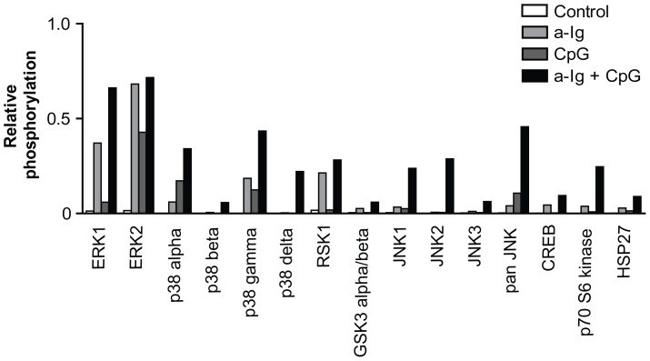
Figure 1. Screening of BCR and TLR9 stimulated kinase activation pattern in BJAB Burkitt's lymphoma cells by MAPK protein profiler array.
BJAB cells were treated with 5 µg/ml anti-Ig and/or 2 µg/ml CpG for 30 minutes. Relative phosphorylation values were calculated as the ratios of signal strengths for each kinases as compared to positive control spots. Only kinases that were activated are shown.

