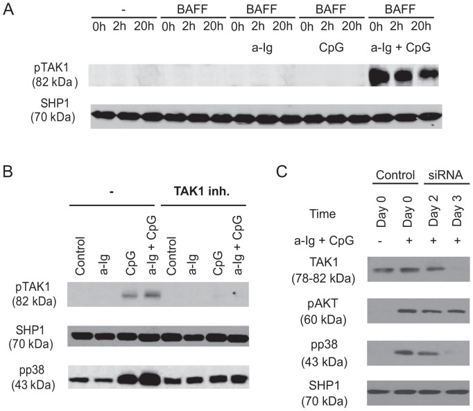
Figure 2. BCR and TLR9 induced signals synergistically activate TAK1 and p38 MAPK in human B cells that is independent of BAFF.
(A) Purified human B cells were left untreated (0 h) or pretreated with 100 ng/ml BAFF for 2 h or 20 h, and then in the last 30 min of pretreatment were activated with anti-Ig (2.5 µg/ml) and/or CpG (1 µg/ml), (B) B cells were stimulated with anti-Ig (2.5 µg/ml), CpG-ODN (2 µg/ml) or the combination of both reagents as indicated for 30 min, in the absence (-) or presence of specific TAK1 inhibitor, (5Z)-7-Oxozeaenol, then samples were subjected to Western blot analysis using pTAK1 or pp38 MAPK specific antibodies. C) Control and TAK1-specific siRNA transfected BJAB cells were activated with 2.5 µg/ml anti-Ig and 1 µg/ml CpG for 30 minutes, and subjected to Western blot analysis to measure TAK1, pAKT and pp38 level. SHP1 was used as a loading control.

