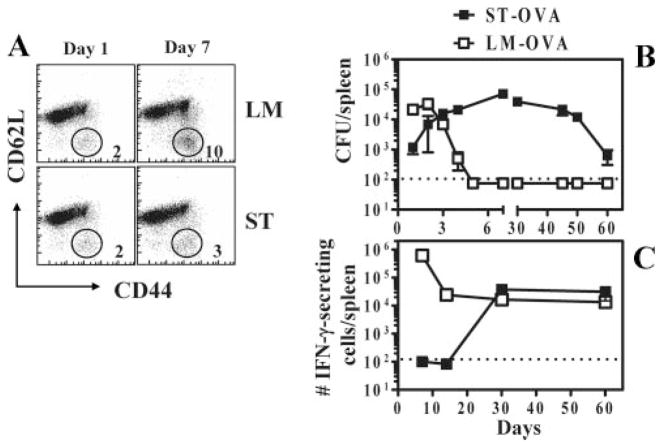
FIGURE 1.
Lack of early T cell activation during infection of mice with ST. B6.129 F1 mice were infected i.v. with 103 LM or ST. A, One and 6 days after infection, spleens were removed from infected mice and stained with anti-CD8, anti-CD44, and anti-CD62L Abs. The relative numbers of activated CD8+ T cells (CD44highCD62Llow) were enumerated. B and C, B6.129 F1 mice were infected i.v. with 103 ST-OVA or LM-OVA. At various time intervals spleens were harvested and the bacterial burden determined (B). Spleens were harvested, processed, and the frequency of OVA-specific CD8+ T cells was enumerated by ELISPOT assay (C). CD8+ T cells were purified using the Dynal magnetic bead system. Purified CD8+ T cells (90–98% pure) were incubated for 48 h with IL-2 (0.1 ng/ml) in the presence or absence of OVA peptide (5 μg/ml), using IP plates (Millipore) precoated with anti-IFN-γ Ab (R46A2; 13 μg/ml in NaHCO3 buffer). Plates were washed with 0.01% PBS with Tween 20 and incubated with biotinylated anti-IFN-γ Ab (XMG1.2). Subsequently the plates were washed, incubated with SA-HRP, and finally developed using 3-amino-9-ethylcarbazole substrate. Dots corresponding to IFN-γ-producing cells were enumerated under a microscope.

