
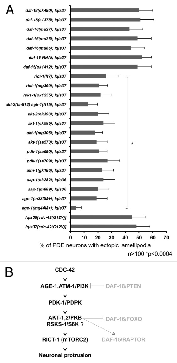
Figure 4. CDC-42 utilizes specific components of the PI3K pathway to drive ectopic lamellipodia formation in PDE neurons. (A) PI3K signaling mutants suppress CDC-42(G12V). The Y-axis denotes the genotype and the X-axis represents the percentage of ectopic lamellipodia formation. “M+” denotes that the animals had wild-type maternal contribution. The number of axons scored > 100. *p < 0.0004 as determined by Fisher Exact Analysis. The error bars represent 2x SEP. (B) A diagram of the genetic interaction with CDC-42 and the PI3K signaling pathway. Molecules in gray are those not involved in CDC-42-induced neuronal protrusion.