
Figure 1.
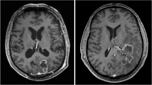
Glioblastoma on T1-weighted, post-contrast magnetic resonance images on the axial view. Tumor localization in the cortex without subventricular zone involvement (left). Glioblastoma with infiltration of the subventricular zone and subependymal spread (right).