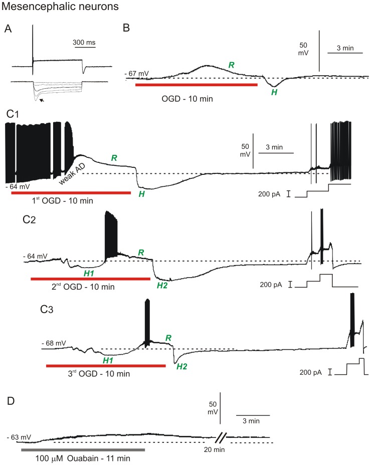
Figure 5. MES neurons strongly resist depolarization in response to OGD or ouabain.
A) MES neurons typically show strong spike accommodation in response to a depolarizing current step (top trace) and strong inward rectification (arrow) in response to a hyperpolarizing current step (bottom trace). B) Whole-cell recording of MES neuron in response to 10 minutes of OGD. The neuron weakly depolarizes followed by active repolarization (R) during OGD. With return to control aCSF, the neuron displays a hyperpolarizing undershoot (H) and returns to near resting potential. C) MES neuron response to three 10-minute OGD exposures. C1) The neuron initially fires spontaneously and inactivates followed by active repolarization during OGD (R). With return of control aCSF, the neuron displays a hyperpolarizing undershoot as in B, before regaining its original membrane potential. At the end of the recording, steps of depolarizing current demonstrate ability to discharge. C2) In response to a second OGD period, the MES neuron initially hyperpolarizes (H1) before firing action potentials that inactivate as in C1, followed by active repolarization during OGD (R). Upon return to control aCSF, the cell hyperpolarizes (H2). Incremental current steps (right) show firing and strong accommodation. C3) As above, the neuron initially hyperpolarizes, inactivates and actively repolarizes during OGD, followed by a brief hyperpolarizing undershoot upon return to control aCSF. D) MES neuron response to 10 minutes ouabain exposure. The cell depolarizes but unlike with OGD, there is no repolarization or hyperpolarizing undershoot upon return to control aCSF.

