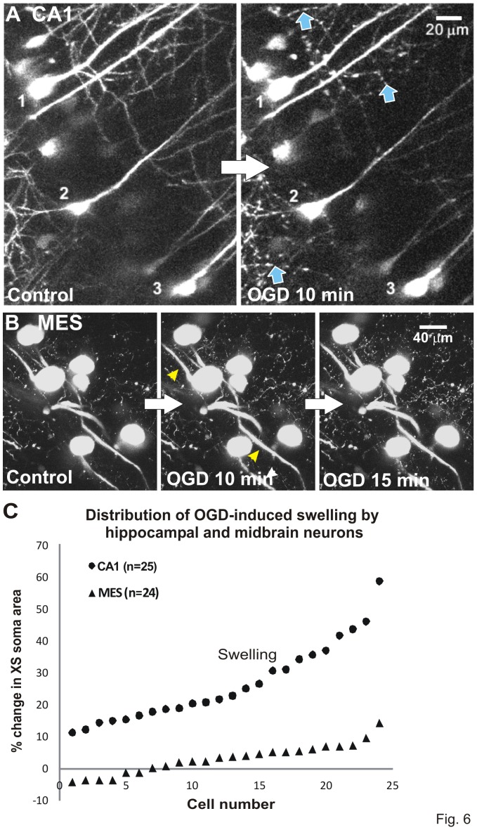
Figure 6. OGD injures CA1 pyramidal neurons but not MES neurons.
Volume responses to OGD by YFP-positive neurons monitored in real time with 2-photon laser microscopy. A) Three CA1 pyramidal cell bodies display swelling as their dendrites form beads (arrows). This is an all-or-none response post-AD [30] and was not further quantified. Both responses denote acute damage caused by 10 minutes OGD. There is no recovery (not shown). B) Neuronal cell bodies in the MES nucleus resist swelling after 10 or 15 minutes of OGD. The primary processes display minor swelling by 10 minutes (arrowheads). C) Change in cross-sectional (XS) area of the cell body is measured to determine soma swelling or shrinking in real time. Swelling by ‘upper’ CA1 neurons is pronounced compared to the small range displayed by ‘lower’ MES neurons in response to 10 minutes OGD. Results are similar to [26] comparing ‘upper’ neocortical pyramidal neurons and ‘lower’ hypothalamic magnocellular endocrine neurons.

