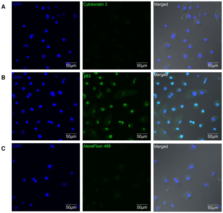
Figure 1. Representative confocal images of immunofluorescence-labelled HCEP cells.
HCEP cells did not express the cornea-specific marker cytokeratin 3 (green) (A) but did express nuclear p63 transcription factor (green) (B). HCEP cells stained with AlexaFluor488 secondary antibody without any primary antibody were served as the negative control (C). Abbreviations: DAPI, 4′,6-diamidino-2-phenylindole.

