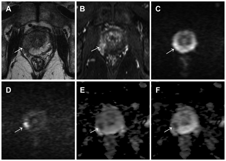
Figure 1. A 77-year-old male with prostate cancer (PSA level of 7.45 ng/mL, Gleason score of 4+3) in the middle right region in the peripheral zone.
Cancer lesion is shown as a homogeneous hypointense lesion with mass effect on T2-weighted imaging (arrow) (A) and focal early enhancement on dynamic contrast-enhanced MR imaging (arrow) (B). The lesion with a focal hyperintensity is depicted clearly in the DW image with b values of 0 and 2000 s/mm2 (arrow) (D), as compared with the DW image with b values of 0 and 1000 s/mm2 (arrow) (C). However, the lesion conspicuity with a focal hypointensity in the ADC map with b values of 0 and 2000 s/mm2 (arrow) (F) was equivalent to that of ADC map with b values of 0 and 1000 s/mm2 (arrow) (E).

