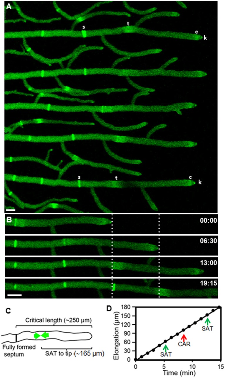
Figure 1. Septum development in hyphae of Neurospora crassa visualized by fluorescent tagging of actin with Lifeact-GFP.
(A) Growth of primary hyphae monitored for up to 20 min. Lifeact-GFP fluorescence reveals the presence of actin in septa (s) and septal actomyosin tangles (t). Actin is also present in subapical collars (c) and the Spitzenkörper (k). (B) Stages in the septation of a hypha. The dotted lines mark the position of the tip at two consecutive times and, predictably, the place where septation occurred ∼6 min later. (C) Critical hyphal dimensions for septation. As a hypha reaches a critical length of ∼250 µm from the last septation site, a new SAT begins to assemble (green arrows) at the future septation site located at about 165 µm from the tip. (D) Kinetics of hyphal elongation and timing of two SAT and CAR events. Note septum formation did not affect the apical growth rate. Scale Bar = 10 µm.

