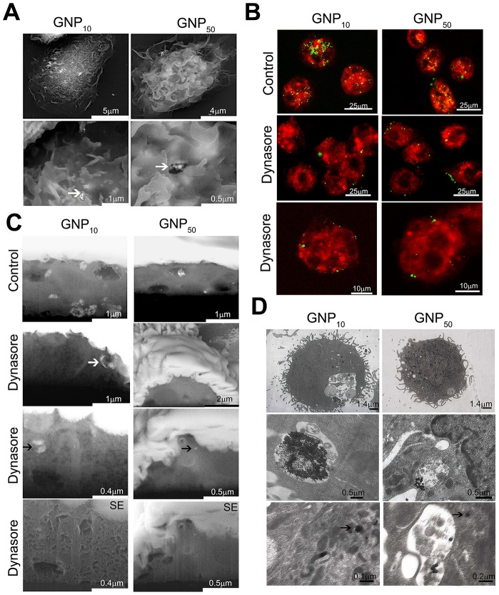
Figure 5. Mechanisms of GNPs internalization by DCs.
The internalization was analyzed by (A) SEM, under different magnifications; white arrows point to GNPs-membrane interactions; (B) Confocal microscopy of live cells stained with FM4-64; or (C) FIB/SEM using BSE, or SE detectors where indicated. White arrow points to the GNP10-loaded vesicle attached to the outer membrane, whereas black arrows point to intracellular presence of GNP10 agglomerates and a single particle of GNP50. The cells shown in (B) and (C) were cultivated with GNPs for 3 h in the presence or absence of Dynasore; (D) TEM, after 24 h-cultures under different magnifications. Back arrows indicate intracellular GNP10 agglomerates and a single GNP50 particle outside the endosome.

