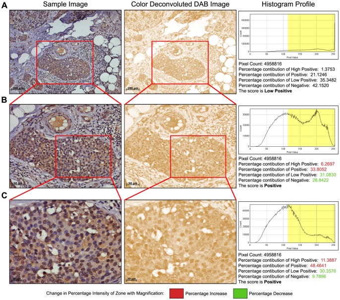
Figure 4. Impact of magnification on image scoring.
A: Analysis of a 10X image area where a significant amount of stroma and fatty tissue is present. After color deconvolution, the score assigned by IHC profiler on the DAB image was determined as low positive. B: Scoring analysis of the same tissue area where image captured was by using a 20X lens in the marked area, focusing more on the actual tumor mass resolute a score of positive. C: Scoring analysis of the same tissue area wherein the image was captured using a 40X lens, focusing more on eliminating the stromal and fatty tissue region increases the percentage of the positive pixels in the positive and high positive zones.

