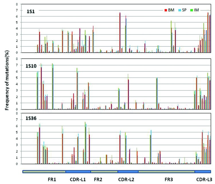
Figure 7. Distribution of somatic mutations along VL: Mutations observed in the three samples along the germline genes 1S1, 1S10 and 1S36. The number of mutations per position from all the reads derived from the same germline gene is plotted as percent of total number of mutations at all the positions for the same set of reads. Mutations for the first 7 positions are not calculated since these positions were encoded by the primer used in V-region amplification.

An official website of the United States government
Here's how you know
Official websites use .gov
A
.gov website belongs to an official
government organization in the United States.
Secure .gov websites use HTTPS
A lock (
) or https:// means you've safely
connected to the .gov website. Share sensitive
information only on official, secure websites.
