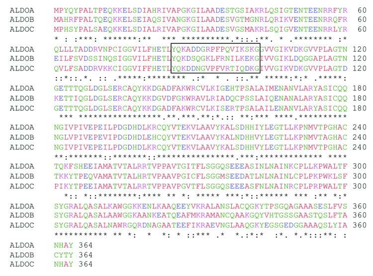
Figure 1. Sequence alignment between the three human aldolase isozymes. Comparison of human aldolase A, B and C amino acid sequences. Sequence 85–102 is boxed. Asterisks (*) represent identical matches in the alignment; colons (:), conserved substitutions; periods (.), semi-conserved substitution. The alignment was performed using the “ClustalW2-Multiple Sequence Alignment” software (www.ebi.ac.uk/Tools/msa/clustalw2).” Red, green, blue and violet correspond to neutral, polar, negative and positive amino acids, respectively.

An official website of the United States government
Here's how you know
Official websites use .gov
A
.gov website belongs to an official
government organization in the United States.
Secure .gov websites use HTTPS
A lock (
) or https:// means you've safely
connected to the .gov website. Share sensitive
information only on official, secure websites.
