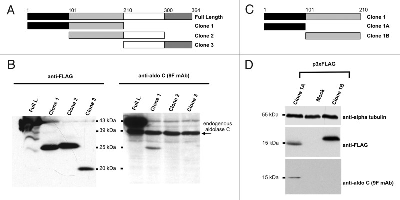
Figure 3. Mapping of the aldolase C region recognized by 9F mAb. (A) and (C) Scheme of the regions of the human aldolase C protein, cloned in the 3xFLAG CMV 7.1 vector. The corresponding clone names are reported on the right. The amino acid limits of each sub-region are indicated by numbers and colors. (B) western blot analysis of total protein lysates from Neuro2a cells alternatively transfected with the 3xFLAG tagged full-length (Full. L.), clone 1, clone 2 or clone 3 fusion products. Proteins were separated by 15% SDS-PAGE and probed with an anti-FLAG antibody (left panel) and with the anti-aldolase C 9F mAb (right panel). (D) western blot analysis of total protein lysates from Neuro2a cells alternatively transfected with the 3xFLAG tagged clone 1A and clone 1B fusion products or mock-transfected. Proteins were separated by 15% SDS-PAGE and probed with an anti-FLAG antibody (upper panel) and with the anti-aldolase C 9F mAb (lower panel). Alpha-tubulin served as loading control.

An official website of the United States government
Here's how you know
Official websites use .gov
A
.gov website belongs to an official
government organization in the United States.
Secure .gov websites use HTTPS
A lock (
) or https:// means you've safely
connected to the .gov website. Share sensitive
information only on official, secure websites.
