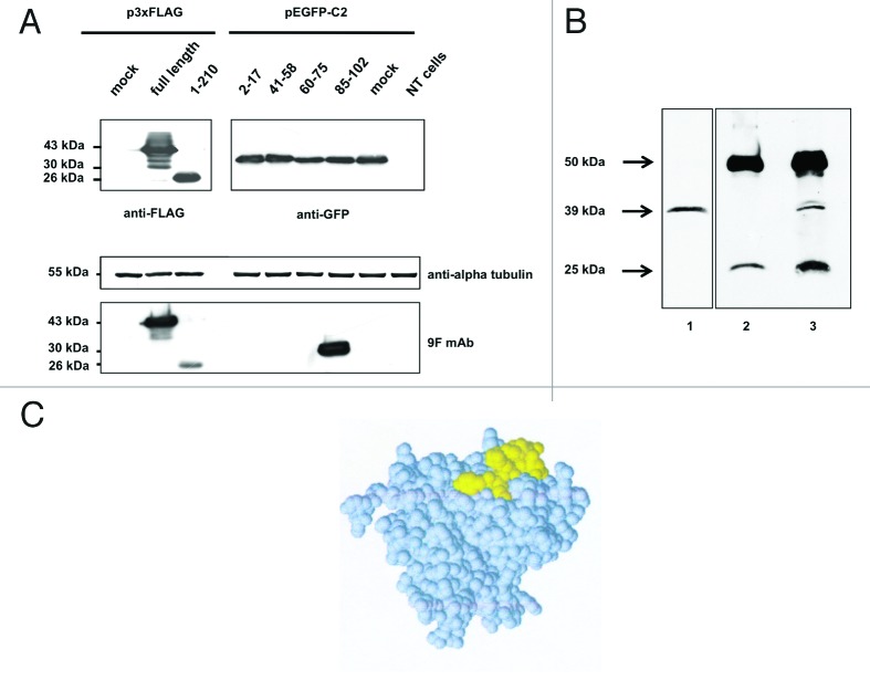
Figure 5. western blot analysis of (A) aldolase C peptides expressed in fusion with a GFP tag at the N-terminus. Total protein extracts obtained from Neuro2a cells alternatively transfected with the four pEGFP-C2 recombinant clones (see Table S2 for details), mock transfected or not transfected were separated by 12% SDS-PAGE and probed using the anti-aldolase C 9F mAb. To have a complete overview of the epitope-mapping, the 3xFLAG-tagged full-length aldolase C and clone 1 fusion proteins were included in the immunoblot panel. Alpha-tubulin served as loading control; anti-FLAG and anti-GFP antibodies were used to verify the transfections. (B) Aldolase C immunoprecipitation with the 9F mAb. Mouse brain lysate (1mg) was immunoprecipitated with the anti-aldolase C 9F mAb (lane 3, IP) after pre-clearing with mouse IgGs (lane 2, IgG). 1/10 of the total immunoprecipitated fractions were analyzed by western blot using the same 9F mAb. Twenty μg of total protein extract were included in the panel as input control (lane 1, Input). (C) Epitope-containing peptide 85–102 mapped on the protein subunit structure. Space filling representation of the aldolase C 3D structure (PDB code 1XFB). Peptide 85–102, which is highly exposed on the protein surface, is shown in yellow.

An official website of the United States government
Here's how you know
Official websites use .gov
A
.gov website belongs to an official
government organization in the United States.
Secure .gov websites use HTTPS
A lock (
) or https:// means you've safely
connected to the .gov website. Share sensitive
information only on official, secure websites.
