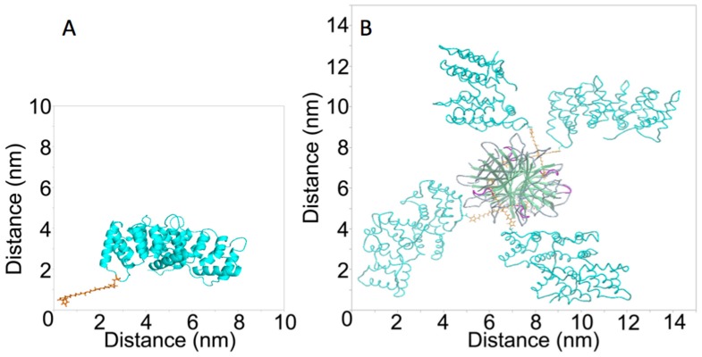
Figure 2. Three-dimensional (3-D) models of anxA5-Biotin (A) and anxA5-NP (B).
The 3-D structure was retrieved from the 1ANX entry of the Protein Data Bank (PDB). Residue 166 was replaced by a cysteine and coupled to maleimide-PEG2-Biotin using Yasara. The picture was generated using PyMOL. The structure of streptavidin was retrieved from the 1SWG entry of PDB. 4 anxA5-biotin monomers were docked to streptavidin’s biotin binding pockets and the picture was generated using ICM-Pro.

