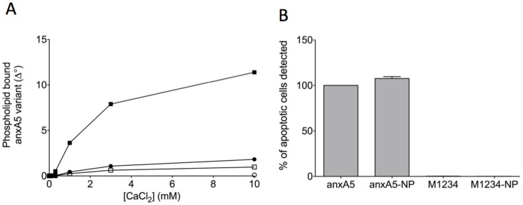
Figure 3. Binding of annexin-variants to phosphatidylserine containing membranes.
Panel A shows calcium-dependent binding of anxA5 (closed circles), M1234 (open circle), anxA5-NP (closed squares) and M1234-NP (open squares) to a synthetic phospholipid surface comprising 20 mole% phosphatidylserine and 80 mole% phosphatidylcholine. Binding was measured by ellipsometry and is expressed as change in degree of the analyser (Δ°) as described elsewhere [24]. Panel B shows the % of apoptotic cells of a population of anti-Fas stimulated Jurkat cells that can be detected using fluorescently labeled annexin-variants and flow cytometry.

