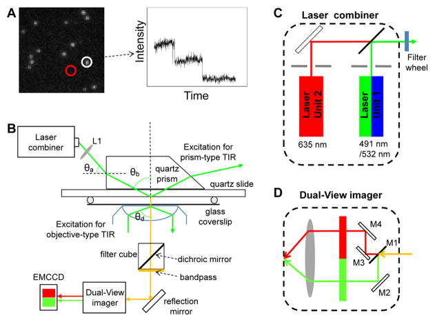
Fig. 1. Single molecule photobleaching assay for direct counting of biomolecules.
(A) Left: A typical initial fluorescence image of individual biocomplexes containing multiple copies of fluorescently labeled molecules prior to photobleaching process. Right: Photobleaching trace of average fluorescent intensity vs. time for the fluorescent spot circled in the image on the left. (B) Schematic of the single molecule TIRF microscope. Adapted from [18] © 2007 with permission from Nature Publishing Group. (C–D) Schematic designs of the optics inside the (C) laser combiner and the (D) Dual-View Imager™. Adapted from [31] © 2010 with permission from Springer.

