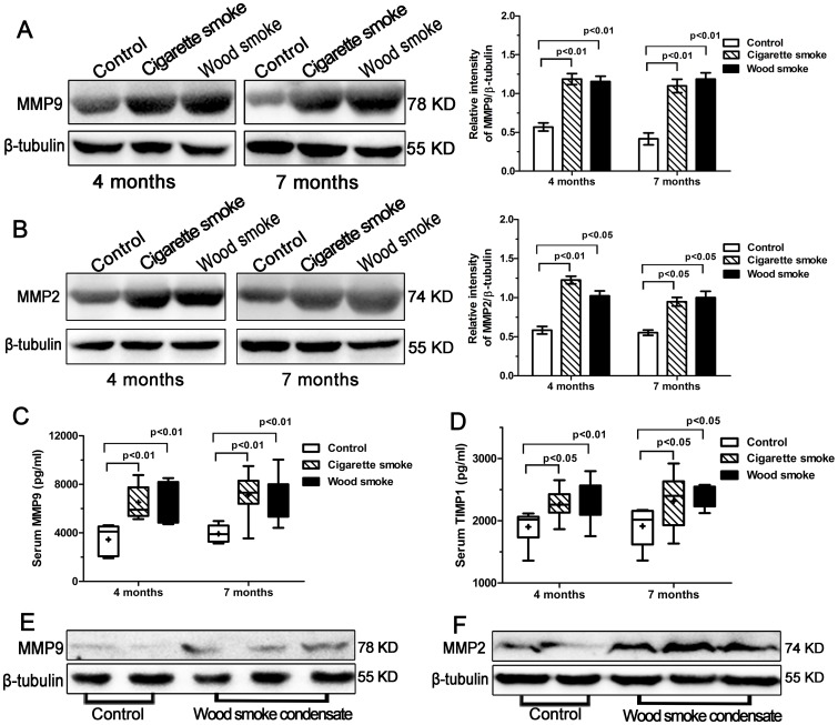
Figure 6. The increased expression of MMP9, MMP2 and TIMP1 by Western blotting or ELISA.
Western blot analysis of MMP9 protein expression (78 KD) in the lung tissues from each group is shown in (A); MMP2 protein expression (74 KD) in (B). Graph showed that the relative intensity of MMP9/β-tubulin protein expression or MMP2/β-tubulin protein expression was higher in lung tissues after smoke exposure for 4 to 7 months compared to controls. (C, D) The levels of serum MMP9 and TIMP1 were significantly higher in the WS group and the CS group than in controls. (E, F) Wood smoke condensate induced an increased expression of MMP9 and MMP2 proteins in primary rat tracheal epithelial cells by Western blotting. Data are shown as the mean ± SEM or as box and whisker plots with the median, minimum and maximum values. n = 8 animals/group.

