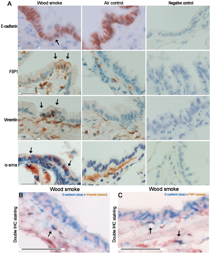
Figure 8. Expression of EMT Markers in Small Airway Walls Exposed to wood smoke.
(A) Photomicrographs showing that the small airways immunostained for E-cadherin, FSP1, vimentin and α-SMA. Although the airway epithelium in rats exposed to WS did not show a marked decrease in E-cadherin immunostaining compared to controls, the positive staining of mesenchymal markers (FSP1, vimentin, α-SMA) was sometimes observed in the small airway epithelium at 7 months. (B, C) Photomicrographs showing the small number of cells that double immunostained for E-cadherin and vimentin or FSP1 in the airway subepithelium of rats exposed to WS for 7 months (only 3 of these 8 rats). Black arrows show positively immunostained cells. Scale bar = 50 µm.

