
Figure 4.
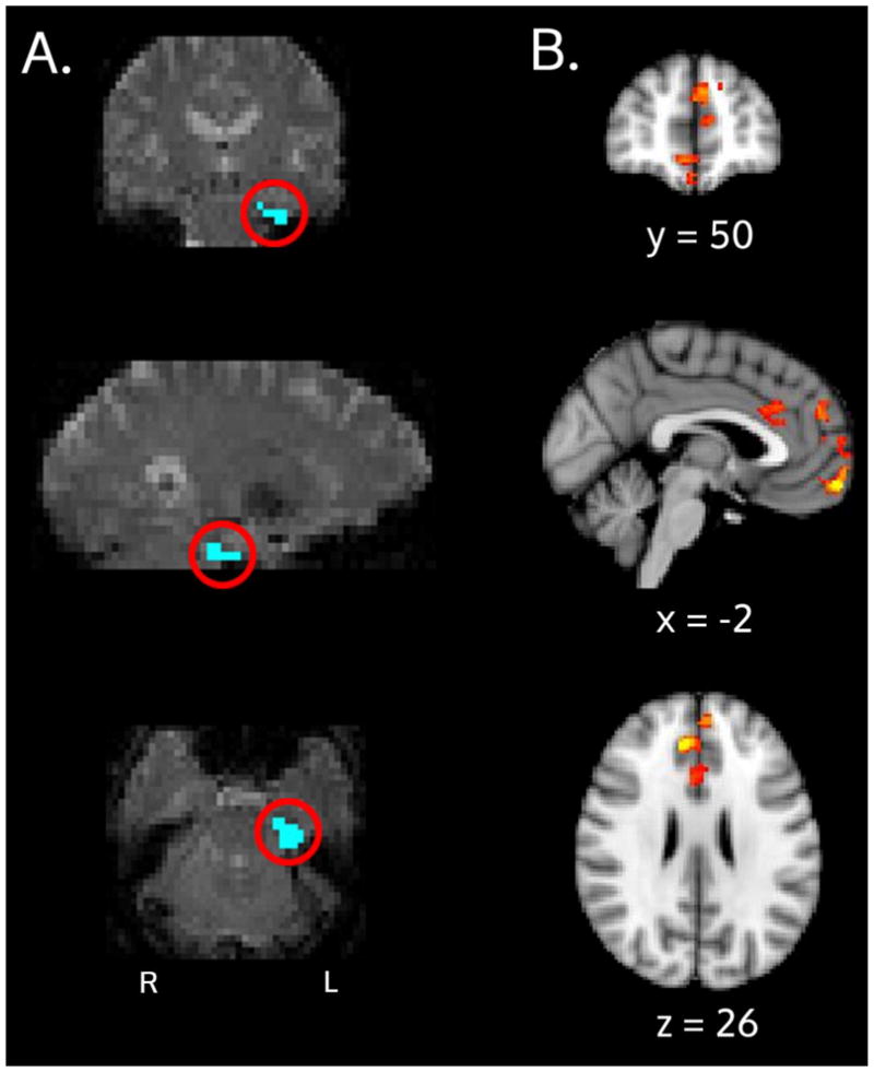
PPI Interaction results: A) The ERC seed (blue, circled in red) for an example subject is shown and overlaid on an individual subject’s fMRI space. B) Group-level results for the APOE x age interaction. These medial prefrontal areas are regions where ε4 carriers showed decreased correlation with the ERC seed and ε3 carriers showed increased correlation, during the retrieval portion of the task.