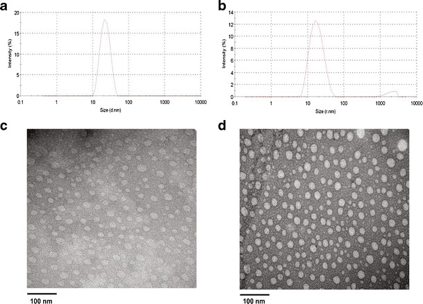
Fig. 2.

Particle size distribution and morphology of curcumin/PEG5K-FTS2 micelles. The average size and size distribution of drug-free micelles (a) and curcumin-loaded PEG5K-FTS2 micelles (b) were examined via DLS analysis. The morphology of drug-free micelles (c) and curcumin-loaded PEG5K-FTS2 micelles (d) was examined via EM following negative staining
