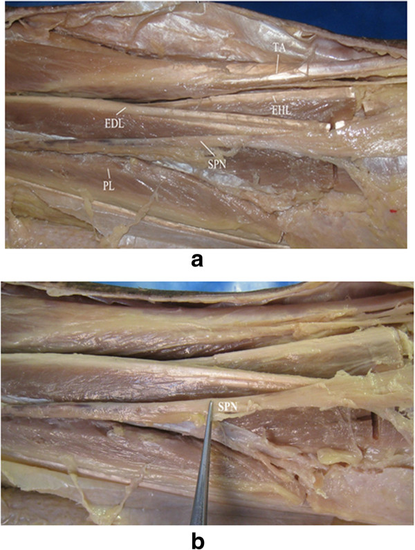
Figure 2.
The superficial structure and the location of SPN. (a) In the superficial structure, each muscle of the anterior compartment was dissected, including tibialis anterior (TA), extensor hallucis longus (EHL), extensor digitorum longus (EDL), and the peroneus longus (PL) and brevis. (b) The location of superficial peroneal nerve (SPN).

