
Figure 1.
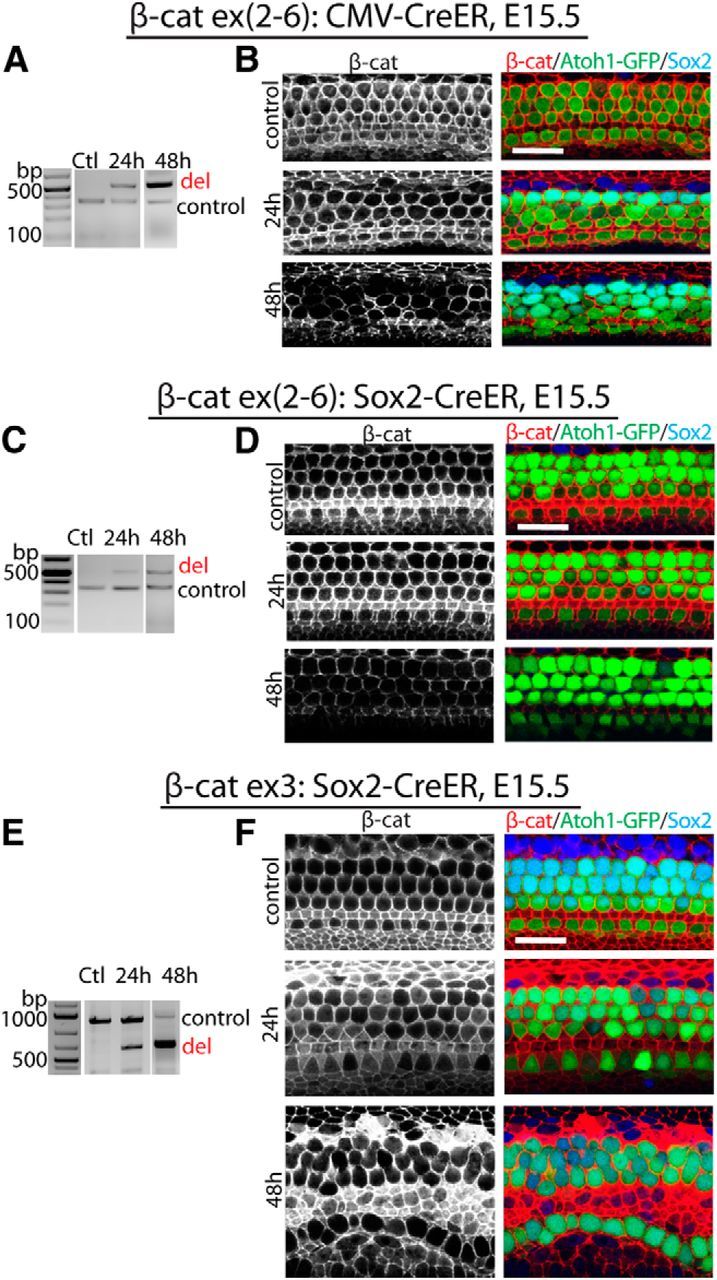
Manipulation of β-catenin expression in developing sensory epithelium. A, In CMV-CreER;β-cateninflox(exon2–6 mutants, exons 2–6 of β-catenin were deleted in sensory epithelium 24 and 48 h after the first dose of tamoxifen at E15.5. B, The decrease of β-catenin expression in sensory epithelium was at its highest point 48 h after the initial dose of tamoxifen. Hair cells were positive for GFP (compound mutants also expressed GFP under the control of Atoh1); β-catenin (red) was expressed in the entire sensory epithelium; Sox2 (blue) was expressed in supporting cells. C, In Sox2-CreER;β-cateninflox(exon2–6) mutants, exons 2–6 of β-catenin were deleted in sensory epithelium 24 and 48 h after the first dose of tamoxifen. D, β-catenin expression in sensory epithelium decreased 48 h after the first dose of tamoxifen. E, Following the initial tamoxifen injection at E15.5 in Sox2-CreER;β-cateninflox(exon3) mutants, deletion of exon 3 of β-catenin was seen at 24 and 48 h in sensory epithelium. F, A greater increase in β-catenin was apparent in sensory epithelium 48 h after the first dose of tamoxifen. The controls are littermates without Cre expression at 48 h after tamoxifen. Scale bar, 20 μm.