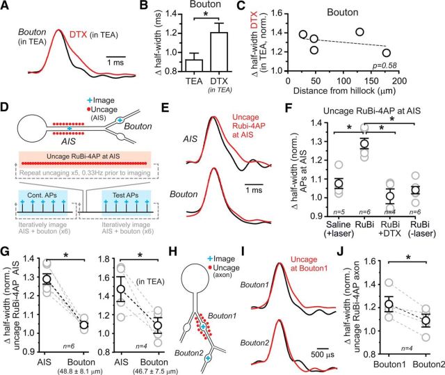
Figure 3.
Local determination of AP repolarization in axons. A, APs recorded at a bouton in a background of 500 μm TEA (black) and following application of 100 nm DTX. Traces are the average of many trials and are shown normalized to the peak of the AP to facilitate comparison. B, Summary of DTX-induced AP widening in TEA at boutons. Data are mean ± SEM; *p < 0.05 by unpaired t test. C, In a background of TEA, lack of dependence of DTX-induced AP widening at a bouton with distance from the axon hillock. Fit of the linear regression is indicated by the dashed line. D, Diagram depicting the experimental configuration. AP recording sites included both the AIS and an axonal bouton located in the same field of view to eliminate need for objective refocusing. Local photolysis of RuBi-4AP (bath applied at 150 μm) was limited to the AIS. Uncaging pulse trains (highlighted in red) were repeated five times at 0.33 Hz before each imaging trial. An imaging trial (highlighted in blue) consisted of five APs stimulated at 0.33 Hz. Measurements of AP waveform were made iteratively between these two sites in control and following RuBi-4AP uncaging. E, APs recorded at the AIS in control (black) and immediately following local photolysis of RuBi-4AP at the AIS. In the same cell, APs were also recorded at a bouton following RuBi-4AP photolysis at the AIS. F, Effect of AIS-directed RuBi-4AP photolysis on AP duration for spikes recorded at the AIS. In addition, control experiments show that laser pulses alone have no effect on AP repolarization and that RuBi-4AP has no basal effect on AP duration without laser-induced uncaging. Data are mean ± SEM; *p < 0.05 by one-way ANOVA. G, Effect of AIS-directed RuBi-4AP photolysis on AP repolarization at the AIS and boutons in basal conditions and in a background of TEA (500 μm). Data are mean ± SEM; *p < 0.05 by paired t test. The distance of the bouton recording position relative to the hillock is indicated below in parenthesis. H, Recording configuration showing that a short region of an axon branch was targeted for local Rubi-4AP photolysis. VSD responses were recorded in alternating trials from boutons located in either the targeted region or on a distal axon branch. I, APs recorded in control (black) and following Rubi-4AP photolysis (red) at bouton locations. J, AP widening, induced at target boutons by Rubi-4AP photolysis, was highly reduced in boutons located on distal axon branches. Data are mean ± SEM; *p < 0.05 by paired t test.

