
Figure 3.
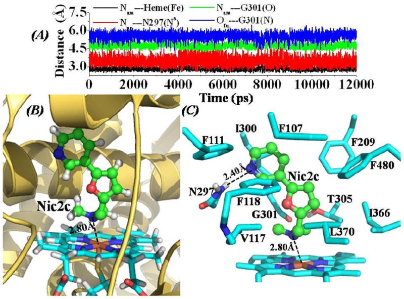
(A) Tracked distances along the MD trajectory for the CYP2A6-Nic2c binding structure. Nam---Heme(Fe) represents the distance between the nitrogen atom on the amine group of Nic2c and the heme iron atom; Npy---N297(Nδ) represents the distance between the nitrogen atom on the pyridine group of Nic2c and the Nδ atom of the N297 side chain; Nam---G301(O) is the distance between the nitrogen atom on the amine group of Nic2c and the carbonyl oxygen atom on the backbone of residue G310; and Ofu---G301(N) represents the distance between the furan oxygen atom of Nic2c and the backbone nitrogen atom of residue G301. (B) QM/MM-optimized CYP2A6-Nic2c binding structure (optimized at B3LYP/6-31G*:Amber8 level). The binding structure is represented in the same color scheme as that in Figure 1A. The dashed line represents the averaged distance between the amine nitrogen atom of Nic2c and the heme iron atom based on the 10 QM/MM optimized structures. (C) Intermolecular interactions in the optimized CYP2A6-Nic2c binding structure. Residues from CYP2A6 within 5 Å of Nic2c are shown in stick style. The averaged distance (based on the 10 QM/MM optimized structures) between the pyridine nitrogen atom of Nic2c and the Hδ atom of N297 side chain is represented as dashed line.