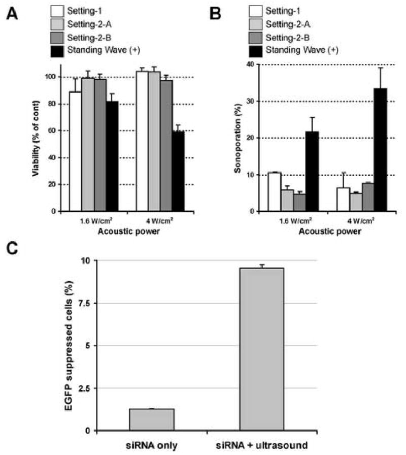
Fig. 4. The impact of standing wave on sonoporation and inhibition of EGFP stable expression in C166-GFP cells by microbubble-enhanced ultrasound delivered siRNA.

A and B, Sonoporation without the presence of standing wave. The concentration of OPTISON was 5% and the exposure time was fixed to 15 sec. Note that both loss of cell viability (A) and sonoporation efficacy (B) have dramatically dropped when standing wave is absent. All data are presented as mean±SD of 3 independent experiments.
C, EGFP gene suppression with intracellular siRNA delivery. The conditions were 5% OPTISON with 1.6 W/cm2 AP × 15 sec exposure and the cells were cultured in a monolyaer fashion. The concentration of siRNA was the same as previously reported where cells were sonicated in suspesion [30]. Approximately half of the sonoporated cells underwent gene supression (mean±SD of 3 independent experiments).
