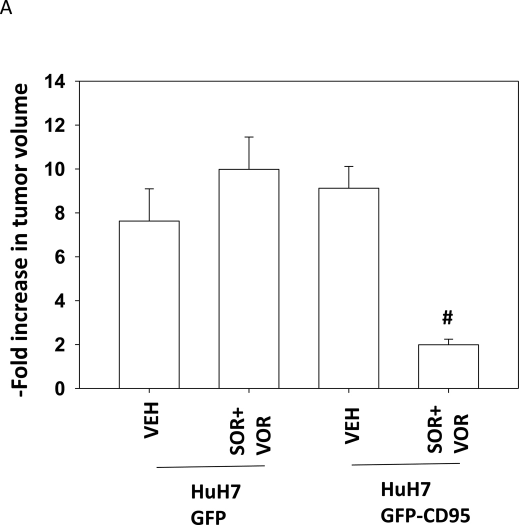
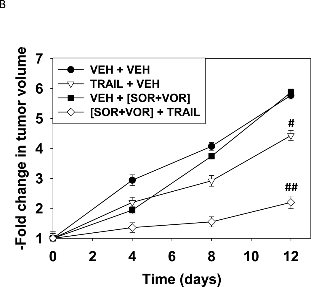
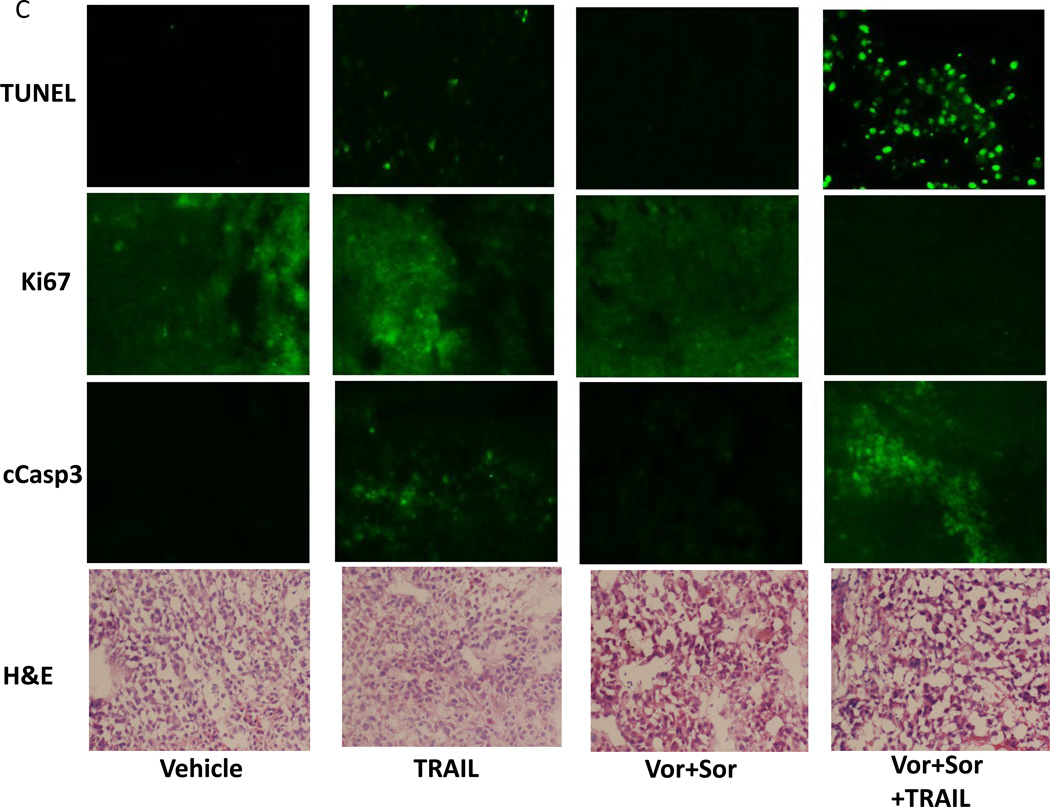
Figure 5. Expression of CD95 enhances the anti-tumor effect of sorafenib and vorinostat in vivo.
(A) HuH7 cells were transfected to express GFP or GFP-CD95. Cells (1×10⁶) were injected into the rear flank of athymic mice. Seven days after injection, prior to obvious tumor formation, animals were treated with vehicle diluent (DMSO, Cremophore) or sorafenib (25 mg/kg) and vorinostat (25 mg/kg) for 5 days. Tumors were permitted to form and 30 days after injection the volume of tumors determined (n =6 animals from 2 independent studies +/− SEM). #p < 0.05 value less than in GFP transfected cells treated with SOR+VOR. (B) HuH7 cells (1×10⁶) were injected into the rear flank of athymic mice. Tumors were permitted to form and 30 days after injection the volume of tumors determined. Initial tumor volumes for each condition were VEH+VEH (223 +/− 55 mm3); SOR+VOR+VEH (229 +/− 69 mm3); VEH+TRAIL (225 +/− 55 mm3); SOR+VOR+TRAIL (227 +/− 53 mm3). Animals were treated with vehicle diluent (DMSO, Cremophore); sorafenib (25 mg/kg) and vorinostat (25 mg/kg); TRAIL (1 mg/kg); and sorafenib and vorinostat and TRAIL for 5 days.(n =6 animals from 2 independent studies +/− SEM). #p < 0.05 value less than in vehicle treated cells; ##p < 0.05 less than TRAIL treated cells. (C) Tumors were removed, fixed and 10 µm slices obtained. Tumor sections were then blocked and subjected to immunohistochemical analysis as per the instructions of the manufacturer for each primary antibody (Ki67 and cleaved caspase-3). The tissue sections were dehydrated, cleared, and mounted with cover slips using Permount.


