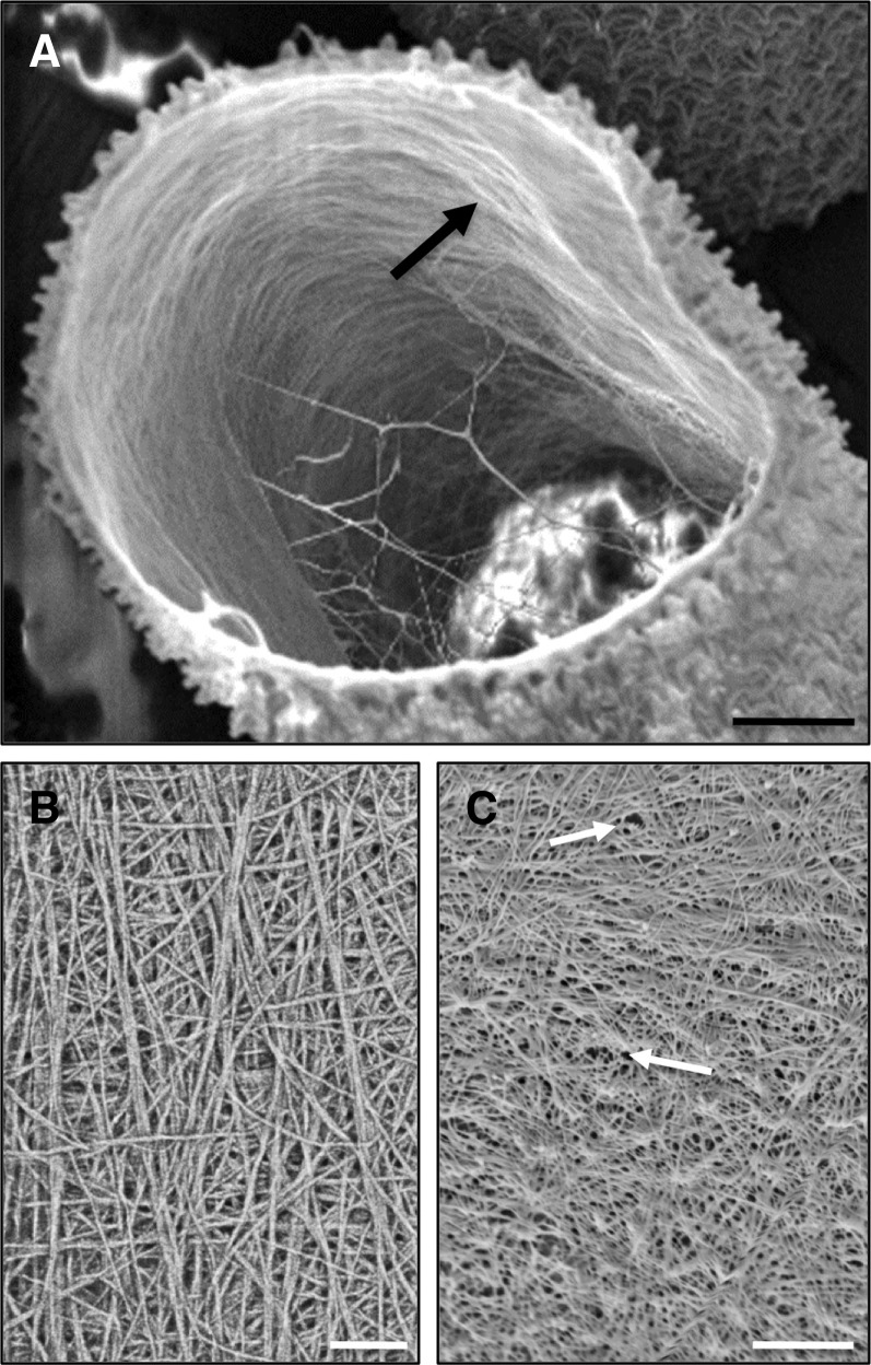
Figure 3.
The inner cellulosic layer of P. margaritaceum. A, Fractured cell showing microfibrils on the innermost layer that are mostly aligned perpendicular to the long axis of the cell. B, Network of cellulose microfibrils showing considerable crosshatching. C, Pores distributed throughout the inner wall layer (arrows). All images were made with FESEM. Bars = 2.8 µm (A), 100 nm (B), and 120 nm (C).

