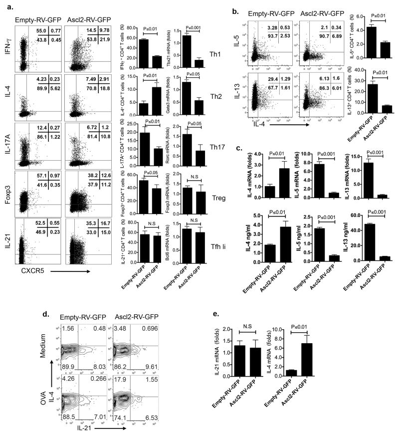
Extended Data Figure 3. Regulation of Th cell differentiation by Ascl2.
a. Naïve CD4+ T cells from C57BL6 mice were activated under neutral condition and infected with Ascl2-RV-GFP or control vector (empty-RV-GFP) virus, followed with continuous culture under Th1, Th2, Th17, iTreg, and Tfh-like condition for 3–4 days, respectively. Quantification of signature genes by intracellular staining and real-time RT-PCR.
(b–c). Ascl2-RV-GFP- or control vector- transduced T cells were cultured under Th2 condition for 4 days.
b. After re-stimulation with PMA and ionomycin for 5h, Th2-related genes including IL-4, IL-5, and IL-13 expression were measured by flow cytometric analysis. c. GFP+ T cells were sorted and re-stimulated by plate-bound anti-CD3, transcriptional expression of IL-4, IL-5, and IL-13 were measured by quantitative RT-PCR; cytokines in supernatants of re-stimulation were subject to ELISA analysis.
(d–e). Ascl2-RV-GFP- or control vector- transduced OT-II cells were adoptively transferred into naïve congenic mice, respectively, followed with subcutaneous OVA/CFA immunization for seven days. d. After restimulation with OVA, flow cytometry analysis of donor-derived cells from dLNs with intracellular IL-4 and IL-21 staining. e. GFP+ donor-derived T cells were sorted from dLNs, re-stimulated with anti-CD3, and subject to quantitative RT-PCR measurement of IL-21 and IL-4 mRNA expression. All data are a representative of two independent experiments. (a–c, and e) bar graph displayed as mean ± SD, n = 3, two-tailed t-test, N.S, no significance.

