
FIG. 2.
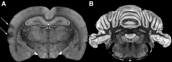
Representative anatomical image data from a double blast exposed animal. Coronal slices through the dorsal hippocampus (A) and the deep cerebellar nuclei (B) are shown. Five of eight double blast exposed animals had cortical contusions of varying sizes, with and without associated subdural hemorrhage (arrows). No gross pathology was visible in the cerebellar white matter.