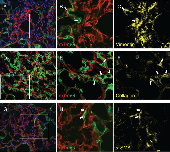
Figure 2.
Detection of epithelial-mesenchymal transition (EMT) in lung sections of Nkx2.1-Cre;mT/mG reporter mice 21 days post-bleomycin instillation. Confocal images demonstrate co-localization of membrane-associated GFP with mesenchymal markers vimentin (A–C), type I collagen (D–F), and α-SMA (G–I). Panels A, D and G show direct detection of Tomato and GFP (red and green) in sections counterstained with DAPI (blue). Original magnification x40. Panels B, E and H show magnified views of the rectangles shown in panels A, D and G, respectively, after cropping and re-sizing using Adobe Photoshop. Lung sections were immunostained for vimentin (C), type I collagen (F) and α-SMA (I) as shown in yellow. Arrows point to representative cells which co-express GFP and mesenchymal markers. Thresholds of staining intensity for the three primary antibodies were established using reference sections treated with only secondary antibodies.

