Figure 8.
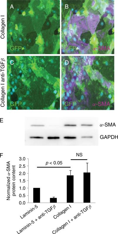
Pan-specific TGFβ neutralizing antibody does not block induction of α-SMA by type I collagen. MAECM grown on type I collagen exhibit irregular and disorganized morphology (A), and a subset of GFP-labeled cells express α-SMA (B; magenta) after 8 days in culture. MAECM grown on type I collagen in the presence of a pan-specific TGFβ neutralizing antibody (2.5 μg/mL) exhibited similar disorganized cell morphology (C) and expression of α-SMA as cells grown on collagen in the absence of neutralizing antibody (D; magenta). Nuclei were counterstained with DAPI (blue). Original magnification x20. E: MAECM treated with and without the pan-specific TGFβ neutralizing antibody were trypsinized from collagen I- and laminin-5-coated polycarbonate filters after 8 days in culture and GFP-positive cells were purified via FACS. Total cell lysates of GFP-positive cells were subjected to SDS-PAGE and immunoblotted with mouse monoclonal anti-α-SMA antibody. Lamin A/C was used as a loading control. Representative Western blot demonstrates that the increase in α-SMA in cells grown on type I collagen is not prevented by the TGFβ neutralizing antibody. F: Relative densitometric values normalized to α-SMA levels in monolayers grown on laminin-5. Results are an average of 3 independent experiments.

