
Figure 2. IMQ induced HIF-1α expression and activation in tumor cells.
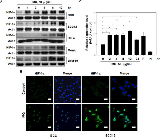
(A) IMQ increased HIF-1α expression in various tumor cell lines. BCC, SCC12, HeLa, MeWo and B16F10 cells were treated with 50 μg/ml IMQ for 0, 1, 2, 4, 8 or 12 hours and then harvested to prepare cell lysates for immunoblotting with HIF-1α and β-actin antibodies. (B) IMQ promoted the nuclear translocation of HIF-1α in tumor cells. BCC and SCC12 cells were treated with or without 50 μg/ml IMQ for 4 hours and then processed for immunocytochemistry using a rabbit anti-HIF-1α antibody and a FITC-conjugated goat anti-rabbit antibody. Scale bars, 20 μm. (C) IMQ enhanced HIF-1α transactivation activity. BCC cells were transfected with a HRE-driven luciferase reporter vector for 48 hours and then treated with 50 μg/ml IMQ for 0, 2, 4, 8, 12 or 24 hours. The cell lysates were collected and processed for a luciferase assay by the detection of luminescence. The P and N groups were the cells transfected with the positive control vector and the negative control vector, respectively. The data are expressed as the mean ± S.E.M. of at least three independent experiments. * p < 0.05; ** p < 0.01.