
Figure 4. The induction of HIF-1α mediated IMQ-induced aerobic glycolysis in tumor cells.
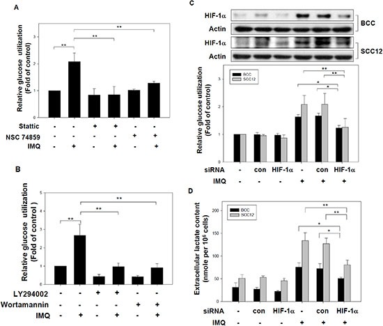
(A) The disruption of STAT3's function inhibited IMQ-induced glucose utilization. BCC cells were incubated in medium containing 10 μM Stattic or 1 μM NSC74859 for 1 hour and then treated with or without 50 μg/ml IMQ for 12 hours. The glucose content assay was performed to determine the relative extracellular glucose content normalized to the total cell number. (B) Inhibition of the PI3K/Akt signaling pathway repressed IMQ-induced glucose utilization. BCC cells were pre-treated with 20 μM LY294002 or 10 μM wortmannin and then treated with or without 50 μg/ml IMQ. The relative glucose utilization level was calculated using a glucose content assay with normalization to the total cell number. (C) and (D) The genetic silencing of HIF-1α suppressed IMQ-induced glucose utilization and lactate production in tumor cells. BCC and SCC12 cells were transfected with a HIF-1α-specific siRNA or a non-specific control siRNA for 24 hours. The transfected cells were treated with 50 μg/ml IMQ. The HIF-1α protein was checked by immunoblotting (up panel of figure C) and the relative glucose utilization level over 12 hours was determined using a glucose content assay (low panel of figure C). The extracellular lactate levels of transfected cells treated with 50 μg/ml IMQ for 24 hours were analyzed using a lactate content assay (D). These results were normalized to the total cell number. The data are expressed as the mean ± S.E.M. of at least three independent experiments. * p < 0.05; ** p < 0.01.