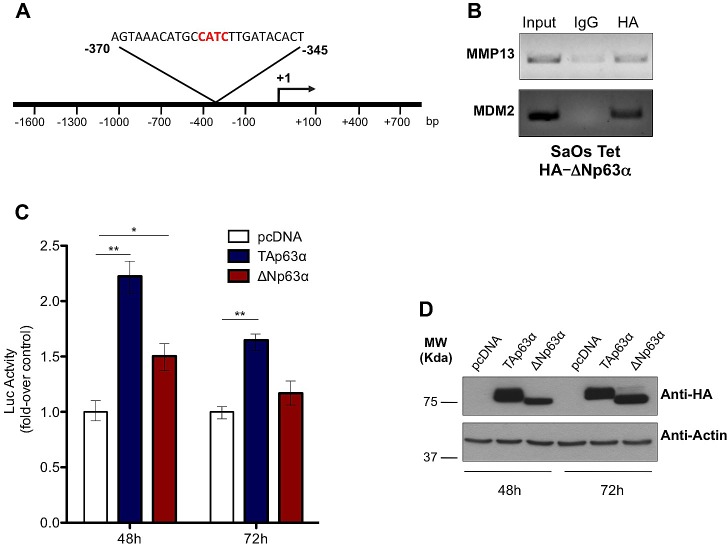
Figure 2. p63 directly binds and transactivates MMP13 promoter.
(a) Schematic map of human MMP13 promoter region with the p53-like RE. The insert shows the sequence of p53 RE, located between -345 and -370 bp upstream of transcription-start site. (b) SaOs-2 cells were induced with 4μg/ml doxycycline for 24h. The sonicated chromatin was bound to ΔNp63α-HA and amplified by PCR with MMP13 primer that recognizes the p53-response element. ChIP on MDM2 promoter was performed as a positive control. Mouse IgG antibody was used as negative control of the ChIP procedure. (c) Both TAp63α and ΔNp63α isoforms transactivate MMP13 promoter at 48h. The MMP13 promoter activity was evaluated after cotransfection with pcDNA vector, TAp63α ΔNp63αplasmids. The luciferase assay was performed after 48h and 72h of cotransfection in 293T cells and was normalized with Renilla luciferase vector. The graphs show a mean ± SD of three different experiments. **, p < 0.01; *, p < 0.05; (d) Western blot analysis on lysates from luciferase assay was performed as control of p63 protein overexpression. Actin was used as loading control.

