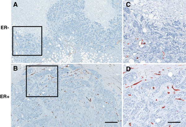
Figure 2.
Representative vasculature images. A) ER- Grade III invasive breast cancer tumor stains against CD34 with as few as 13 quantified vessels at a region adjacent to the tumor edge. Scale = 800 μm. This may be compared with B) ER + CD34 stained grade III tumor which has as many as 84 vessels in the same area as evidenced by C) and D) which are enlarged views of the inset areas with quantified vessels of each masked in red. Scale bar = 200 μm.

