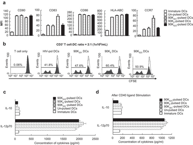
Figure 2.
Characteristics of DCs pulsed with the 90K peptide. (a) FACS was performed to analyze cell surface markers (CD80, CD83, CD86, HLA-ABC and CCR7) of DCs pulsed with or without 90K peptides. Data are expressed as the mean fluorescence intensity increase over the isotype control±s.d. from three independent experiments. (b) Cell proliferation, as measured by CFSE dilution, of CD3+ T cells from a HLA-A*0201+ blood donor in response to unpulsed DCs or DCs pulsed with 90K peptides or the control HIV-pol peptide. Data are expressed as percentages of the mean frequency of dividing CD3+ T cells in three independent experiments. (c, d) Cytokines secreted into the culture supernatants were measured by ELISA. Production of IL-12p70 and IL-10 during maturation of DCs (c) and after stimulation with CD40 ligand-transfected J558 cells (d). The results are illustrated as the mean (pg/ml)±s.d. of triplicate cultures from two independent experiments. CFSE, 5(6)-carboxyfluorescein diacetate succinimidyl ester; DC, dendritic cell; FACS, fluorescence-activated cell sorting analysis; IL, interleukin.

