Figure.

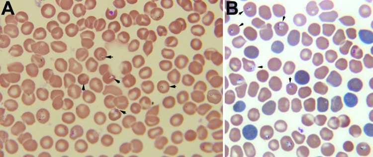
A) Giemsa-stained thin blood smear for an 8-year-old boy from China showing erythrocytes with typical ring forms, paired pyriforms, and tetrads of a Babesia sp. (arrows). B) Giemsa-stained thin blood smear for a mouse with severely combined immunodeficiency, which had been injected with blood from the patient, showing Babesia sp.–infected erythrocytes (arrows). Original magnifications ×1,000.
