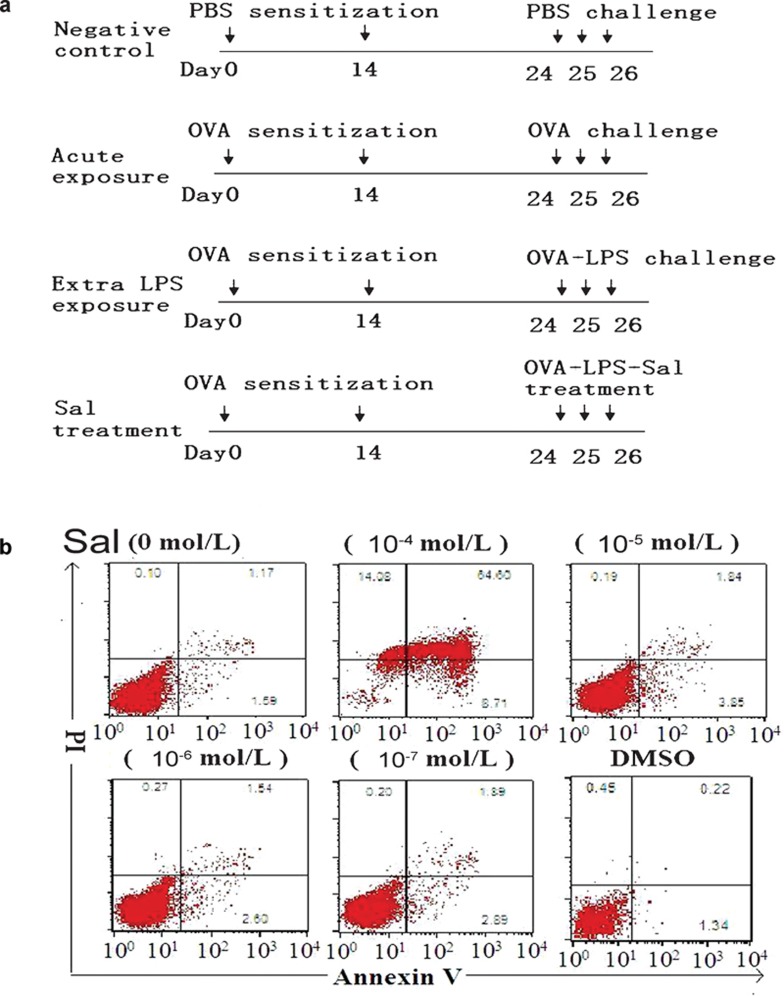
Figure 1.
Experimental design and salmeterol concentration. (a) Experimental protocol. Mice were sensitized with PBS or OVA plus alum on days 0 and 14 and then were challenged with an aerosolized form of different drugs for 3 consecutive days. The extra-LPS exposure group of mice was exposed to 1% OVA and 0.01% LPS aerosol for 3 days. The salmeterol treatment group of mice was exposed to 1% OVA, 0.01% LPS and 10−5 mol/l salmeterol aerosol for 3 days. (b) The highest concentration of salmeterol increases apoptosis in DCs. On day 6, DCs cultured in GM-CSF and IL-4 were treated with different concentrations of salmeterol for 24 h. DCs treated with salmeterol were then harvested and labeled with annexin V/PI. The numbers indicate percentages of PI- or annexin V-positive cells. DC, dendritic cell; GM-CSF, granulocyte/macrophage colony-stimulating factor; LPS, lipopolysaccharide; OVA, ovalbumin; PBS, phosphate-buffered saline; PI, propidium iodide.

