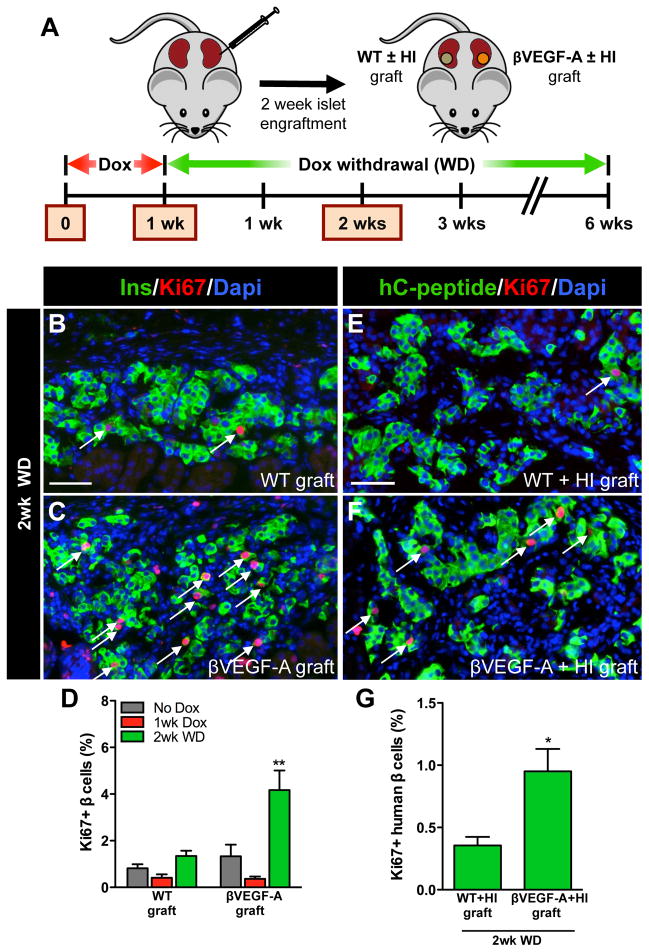
Figure 4. β cell replication is independent of the pancreatic site and soluble circulating factors and is not limited to murine β cells.
(A) Islets from βVEGF-A mice and WT controls were transplanted into βVEGF-A recipients or mixed with human islets (HI) and transplanted into NOD-scid-IL2rγnull mice. Islets engrafted for 2 wks then grafts were harvested and analyzed at No Dox, 1wk Dox, and 2wk WD time points; n=3–4 mice/time point. (B–C) β cell proliferation at 2wk WD in WT and βVEGF-A islet grafts; insulin (Ins, green), Ki67 (red), and Dapi (blue). Scale bar is 50 μm and applies to B–C. (D) Quantification of β cell proliferation in WT and βVEGF-A islet grafts. **, p<0.01, 2wk WD vs. No Dox and 1wk Dox across graft types. (E–F) β cell proliferation at 2wk WD in WT+HI and βVEGF-A+HI grafts; human C-peptide (hC-peptide, green), Ki67 (red), and Dapi (blue). Scale bar is 50 μm and applies to E–F. (G) Quantification of β cell proliferation in WT+HI and βVEGF-A+HI grafts at 2wk WD. *, p<0.05.

