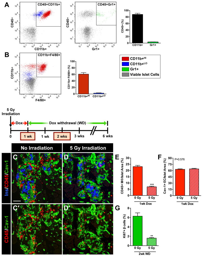
Figure 6. Recruitment of MΦ into βVEGF-A islets upon VEGF-A induction is necessary for the β cell proliferative response.
(A–B) Flow cytometry analysis of βVEGF-A islets after 1-wk Dox treatment. (A) The CD45+ BMC population recruited to βVEGF-A islets was composed of 90% CD11b+ MΦs and 3% Gr1+ cells. (B) F4/80 expression in subsets of more mature CD11b+HI (60%) and less mature CD11b+LO (3%) MΦs; n=5 islet preparations (1–2 mice/preparation). (C–G) Partial bone marrow ablation prior to VEGF-A induction blocks MΦ recruitment and prevents β cell proliferation. (C, D) Immediately after sublethal irradiation, VEGF-A expression in βVEGF-A mice was induced for 1 wk by Dox administration and tissues were examined for the presence of CD45+ MΦs at 1wk Dox and compared with non-irradiated controls; n=4 mice/group; insulin (Ins, blue), CD45 (red) and EC marker caveolin-1 (Cav-1, green). Panel C′ and D′ show CD45 (red) and caveolin-1 (green) labeling. Scale bar is 50 μm and applies to C–D′. (E) Sublethal irradiation reduced infiltration of CD45+ MΦs, ***, p<0.001, 0 Gy vs. 5 Gy. (F) Sublethal irradiation did not affect intra-islet EC expansion, p=0.5760, 0 Gy vs. 5 Gy. (G) Two wks after Dox withdrawal, β cell proliferation was significantly reduced in sublethally irradiated βVEGF-A mice vs. non-irradiated controls, **, p<0.01; n=4 mice/group.

