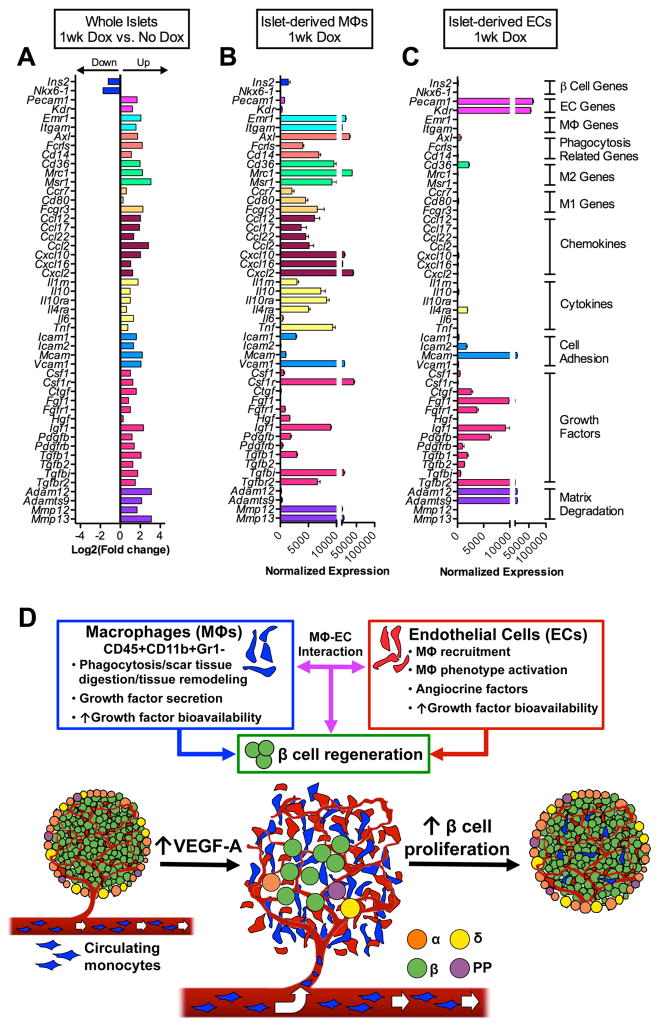
Figure 7. Transcriptome analysis of βVEGF-A islets and purified islet-derived MΦs and ECs.
(A–C) Gene expression profile of whole βVEGF-A islets, islet-derived MΦs, and islet-derived ECs by RNA-Seq; n=3 replicates/each sample set. (A) Differential expression of β cell-, EC-, MΦ-specific genes, phagocytosis-related genes, MΦ phenotype markers (M1, classical; M2, alternative), chemokines, cytokines, cell adhesion molecules, growth factors, and matrix degrading enzymes between islets at 1wk Dox vs. No Dox, p<0.05 for fold change ≥2. See also data in Figure S6M. (B) At 1wk Dox, recruited MΦs are highly enriched for transcripts of phagocytosis-related genes, M2 markers, chemokines, cytokines, cell adhesion molecules, and metalloproteinases involved in tissue repair. (C) Intra-islet ECs mainly express growth factors and matrix degrading enzymes that facilitate growth factor release from the extracellular matrix. Data in B and C are plotted as mean ± SEM. (D) New paradigm for the role of MΦ – EC interactions in β cell regeneration. Upon VEGF-A induction intra-islet ECs proliferate and circulating monocytes recruited to islets differentiate into MΦs. These CD45+CD11b+Gr1− recruited MΦs and ECs produce effector molecules that either directly or cooperatively induce β cell proliferation and regeneration.

