
Figure 4.
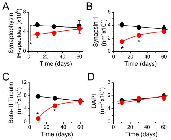
Quantification of immunohistochemical labeling in the NTS. A–D: Stereological quantification of synaptophysin (A), synapsin-1 (B), and β-III tubulin (C) revealed the loss and restoration of synapses and fibers following the vagotomy, without a change in neuronal/glia density in the NTS as shown by DAPI-labeled nuclei (D). *P < 0.01 versus sham controls.