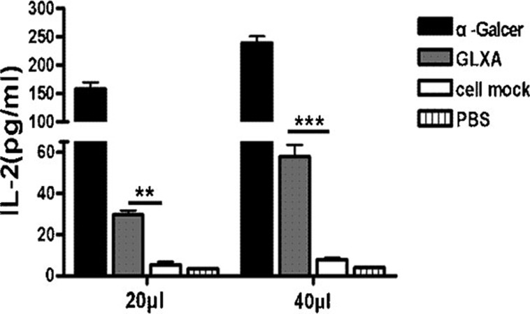
Figure 2.
Recognition of GLXA by iNKT hybridoma cells using cell-free antigen-presentation assay. iNKT hybridoma cells (5×10⁴ cells per well) were added and cultured in CD1-coated 96-well plates in the presence of α-Galcer, GLXA, cell mock or PBS at different doses (20 or 40 µl) for 24 h. The concentrations of α-Galcer in the culture were 0.05 or 0.1 µg/ml, while the concentrations of GLXA were 2.5 or 5 µg/ml. Concentration of IL-2 in supernatant was determined by ELISA. One representative experiment of three independent experiments is shown. The results are shown as mean±s.d. of each group (**P<0.01 and ***P<0.001). α-Galcer, α-galactosylceramide; GLXA, glycolipid exoantigen; iNKT, invariant natural killer T; PBS, phosphate-buffered saline.

