
Figure 5.
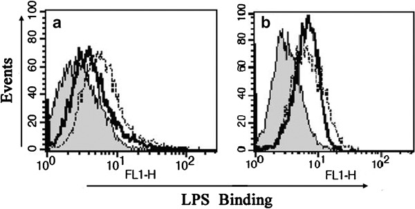
Suppression of the binding of smooth LPS to THP-1 cells by MBL. THP-1 cells were pre-incubated with (black solid line) or without (dotted line) MBL for 30 min (a) or with anti-MBL pAb and MBL (black solid line) for 30 min (b) and then further incubated at 4 °C for 30 min with Alexa488-labeled smooth LPS (E. coli O111:B4). The binding of LPS on the cell surface was determined by FCM. The histograms shown are representatives of three experiments. Shaded curves represent the negative control without labeled LPS. FCM, flow cytometry; LPS, lipopolysaccharide; MBL, mannan-binding lectin; pAb, polyclonal antibody.