
Fig. 3.
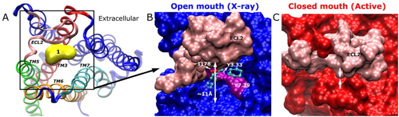
(A) Allosteric Site 1 is located in the extracellular mouth as illustrated in the X-ray structure. The ECL2, TM3, TM5, TM6 and TM7 are highlighted in pink, red, green, orange and cyan, respectively. (B) In the X-ray structure, the M2 receptor exhibits opened extracellular mouth by ∼11 Å as measured between the Cβ atoms of Tyr177ECL2 and Trp4227.35. The receptor is represented by surface with pink for ECL2, purple for key interacting residues and blue for the remainder of the receptor. The bound probes are rendered as ball-and-sticks. (C) In the active conformer (red), ECL2 (pink) is found to close up the extracellular mouth.